


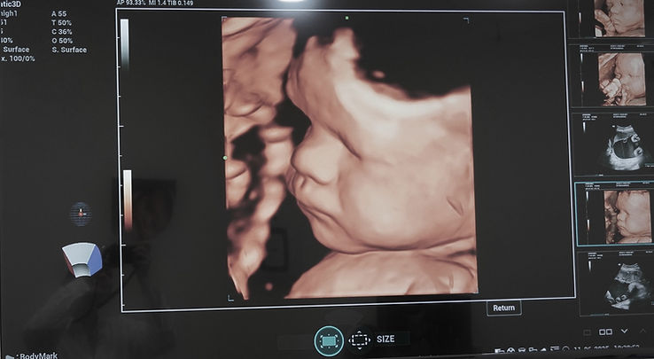
What is Ultrasound?
Sonography is an essential medical imaging tool that utilizes high-frequency sound waves to produce real-time images of the body. As these sound waves reflect off internal anatomy, they create immediate visual data on a display. Because this method uses no ionizing radiation, it is exceptionally safe for patients of all ages.
This non-invasive diagnostic technology allows clinicians to inspect internal structures without making surgical incisions. At GI Rimon, our specialized medical center operates under strict Israeli standards at affordable Georgian prices. We follow rigorous ESHRE and Ministry of Health protocols to ensure every patient receives safe, boutique medical care.
History of Ultrasound Diagnostics
Originally developed from industrial and maritime acoustic navigation, sonogram technology quickly evolved into an indispensable medical tool. Early machines produced basic black-and-white outlines, but decades of continuous engineering have revolutionized diagnostic imaging. Today, modern equipment operates with remarkable clarity and computational speed.
Contemporary clinical systems can analyze blood flow and soft tissues with exceptional fidelity. This historical evolution has transformed ultrasonography from a simple investigative tool into a cornerstone of modern clinical practice. Our clinic continuously upgrades its technology to maintain over 5 years of exceptional results.
Types of Ultrasound
Different scanning modalities exist to serve specific clinical needs across various specialties. Standard three-dimensional and Doppler ultrasound systems evaluate the heart and blood vessels or examine the digestive tract. We also utilize specialized vascular scans to assess circulation without discomfort.
Depending on your symptoms, a physician may recommend targeted pelvic imaging or an abdominal check. Whether evaluating the thyroid, checking liver health, or analyzing a breast lesion, each type of ultrasound provides vital clinical insights. This versatility ensures that every individual or couple receives tailored diagnostic support.
The Ultrasound Examination Procedure
Preparing for the Ultrasound Scan
Proper preparation ensures optimal image quality during your session. For certain scans involving the stomach or liver, you may be asked to fast for several hours beforehand. This prevents bowel gas from obscuring the view of deep abdominal organs.
Conversely, when evaluating the bladder or urinary system, drinking water to fill the bladder is often required before we perform the test. Clear instructions are always provided by our staff prior to your appointment. Proper preparation allows the sonographer to capture the clearest possible pictures.
Step-by-Step Guide to the Examination
During the appointment, the ultrasound technologist applies a warm, hypoallergenic gel directly to the skin over the target area. A handheld transducer or probe is then moved gently across the skin to examine the anatomical region of interest. This gel eliminates air pockets, allowing acoustic waves to travel freely.
As the probe moves, acoustic waves reflect off internal anatomy and send echoes back to the system. The machine instantly processes these signals to display real-time images on the screen. The entire process is conducted with the utmost sensitivity and respect for your comfort.
What to Expect During the Scan
The scanning session is completely painless and typically lasts between 15 and 30 minutes. You will rest comfortably in our modern clinic room while the operator captures effective imaging data. There is no discomfort involved, making it suitable for even the most sensitive patients.
Once the evaluation is complete, the gel is easily wiped away with a clean towel. Because there is no sedation or surgical intervention required, you can immediately return to your normal daily activities. Our team ensures that your experience is calm, supportive, and efficient.