
6010, Georgia, Batumi, Rustaveli Ave, 39
M-F 10-17 | Sat 10-14 | Sun: OFF

Ultrasound Examination & Diagnostics – Scan Procedure and Results

conclusion image

Understanding Ultrasound Examination

What is Ultrasound?

Sonography is an essential medical imaging tool that utilizes high-frequency sound waves to produce real-time images of the body. As these sound waves reflect off internal anatomy, they create immediate visual data on a display. Because this method uses no ionizing radiation, it is exceptionally safe for patients of all ages.
This non-invasive diagnostic technology allows clinicians to inspect internal structures without making surgical incisions. At GI Rimon, our specialized medical center operates under strict Israeli standards at affordable Georgian prices. We follow rigorous ESHRE and Ministry of Health protocols to ensure every patient receives safe, boutique medical care.

History of Ultrasound Diagnostics

Originally developed from industrial and maritime acoustic navigation, sonogram technology quickly evolved into an indispensable medical tool. Early machines produced basic black-and-white outlines, but decades of continuous engineering have revolutionized diagnostic imaging. Today, modern equipment operates with remarkable clarity and computational speed.
Contemporary clinical systems can analyze blood flow and soft tissues with exceptional fidelity. This historical evolution has transformed ultrasonography from a simple investigative tool into a cornerstone of modern clinical practice. Our clinic continuously upgrades its technology to maintain over 5 years of exceptional results.

Types of Ultrasound

Different scanning modalities exist to serve specific clinical needs across various specialties. Standard three-dimensional and Doppler ultrasound systems evaluate the heart and blood vessels or examine the digestive tract. We also utilize specialized vascular scans to assess circulation without discomfort.
Depending on your symptoms, a physician may recommend targeted pelvic imaging or an abdominal check. Whether evaluating the thyroid, checking liver health, or analyzing a breast lesion, each type of ultrasound provides vital clinical insights. This versatility ensures that every individual or couple receives tailored diagnostic support.

The Ultrasound Examination Procedure

Preparing for the Ultrasound Scan

Proper preparation ensures optimal image quality during your session. For certain scans involving the stomach or liver, you may be asked to fast for several hours beforehand. This prevents bowel gas from obscuring the view of deep abdominal organs.
Conversely, when evaluating the bladder or urinary system, drinking water to fill the bladder is often required before we perform the test. Clear instructions are always provided by our staff prior to your appointment. Proper preparation allows the sonographer to capture the clearest possible pictures.

Step-by-Step Guide to the Examination

During the appointment, the ultrasound technologist applies a warm, hypoallergenic gel directly to the skin over the target area. A handheld transducer or probe is then moved gently across the skin to examine the anatomical region of interest. This gel eliminates air pockets, allowing acoustic waves to travel freely.
As the probe moves, acoustic waves reflect off internal anatomy and send echoes back to the system. The machine instantly processes these signals to display real-time images on the screen. The entire process is conducted with the utmost sensitivity and respect for your comfort.

What to Expect During the Scan

The scanning session is completely painless and typically lasts between 15 and 30 minutes. You will rest comfortably in our modern clinic room while the operator captures effective imaging data. There is no discomfort involved, making it suitable for even the most sensitive patients.
Once the evaluation is complete, the gel is easily wiped away with a clean towel. Because there is no sedation or surgical intervention required, you can immediately return to your normal daily activities. Our team ensures that your experience is calm, supportive, and efficient.

Ultrasound Diagnostics: Interpreting Results

Understanding Ultrasound Images

Specialized ultrasound imaging translates returning echoes into a dynamic grayscale picture on the screen. Dense biological materials like bone reflect strong echoes and appear bright white, while fluid-filled areas appear dark or black. A skilled specialist evaluates these visual variations to understand tissue health.
By analyzing these distinct shades and contours, a clinician can easily differentiate between healthy anatomy and a physical abnormality. This immediate clarity helps physicians diagnose complex medical conditions rapidly and safely. Our medical team takes the time to explain these visual findings clearly to every patient.

Common Findings in Ultrasound Diagnostics

Scan evaluations can reveal a wide variety of important clinical details across different body systems. For example, a physician might detect a benign cyst in the kidney or spot small gallstones within the digestive tract. In gynecological care, scans help identify structural issues within the reproductive system.
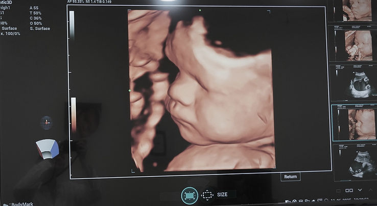
These evaluations are also indispensable during maternity care, allowing doctors to track fetal growth during pregnancy. Whether checking heart and blood vessels or assessing organ function, the findings provide a clear roadmap for your healthcare. Every result is reviewed by experienced specialists to guarantee accuracy.

Follow-Up Procedures After the Examination

If a scan reveals an unexpected finding, your doctor will guide you through the appropriate next steps. In many cases, a routine followup scan is scheduled to monitor how a benign feature behaves over time. This watchful observation ensures that any changes are caught immediately without invasive testing.
When a definitive confirmation is required, sonography provides precise real-time guidance for interventions like a needle biopsy. This targeted approach allows the physician to obtain a tissue sample safely and with minimal discomfort. This step is often essential to establish a conclusive, accurate diagnosis.

Advancements in Ultrasound Technology

Innovations in Ultrasound Scanning

Recent technological leaps have transformed sonography from basic anatomical outlining into high-definition clinical mapping. Modern systems utilize advanced computer processing to evaluate blood flow and inspect internal organs with unprecedented fidelity. These innovations deliver incredible diagnostic clarity without exposing patients to ionizing rays.
Today, high-resolution transducers can visualize subtle microvascular circulation that older machines could never see. This capability allows specialists to identify structural anomalies at their earliest, most treatable stages. Our boutique clinic invests heavily in these cutting-edge innovations to maintain superior clinical standards.

The Future of Ultrasound Diagnostics

The future of medical scanning is being shaped by the integration of artificial intelligence and automated image analysis. Smart software algorithms are now being engineered to enhance image resolution and improve diagnostic accuracy. These digital tools assist clinicians by flagging subtle visual patterns automatically.
In the coming years, these advancements will help doctors detect microscopic changes in any anatomical structure faster than ever before. This synergy of human expertise and advanced computing will make preventive screenings even more dependable. Such progress reinforces sonography as an indispensable pillar of modern healthcare.

Impact on Patient Care and Outcomes

At GI Rimon, our individualized care model leverages these advanced scanning technologies to deliver exceptional medical support. For pregnant women, prenatal sonography offers reassuring views of gestational progress without any physiological risks. This gentle, patient-centered approach ensures complete peace of mind during every consultation.
By operating according to strict Israeli standards at affordable Georgian prices, we make premium healthcare accessible to all. Our commitment to clinical excellence and rapid, reliable reporting consistently improves long-term patient outcomes. We are proud to serve our community with unmatched medical dedication and compassion.


Need a consultation?
Just leave your contact details and we'll reach out to you